Conditions We Treat
Swallowing Disorders
Feeding Tube Dependence
Pharyngoesophageal Stenosis
Upper Esophageal Sphincter Dysfunction
Zenker’s Diverticulum
Esophageal Disorders
Voice Disorders
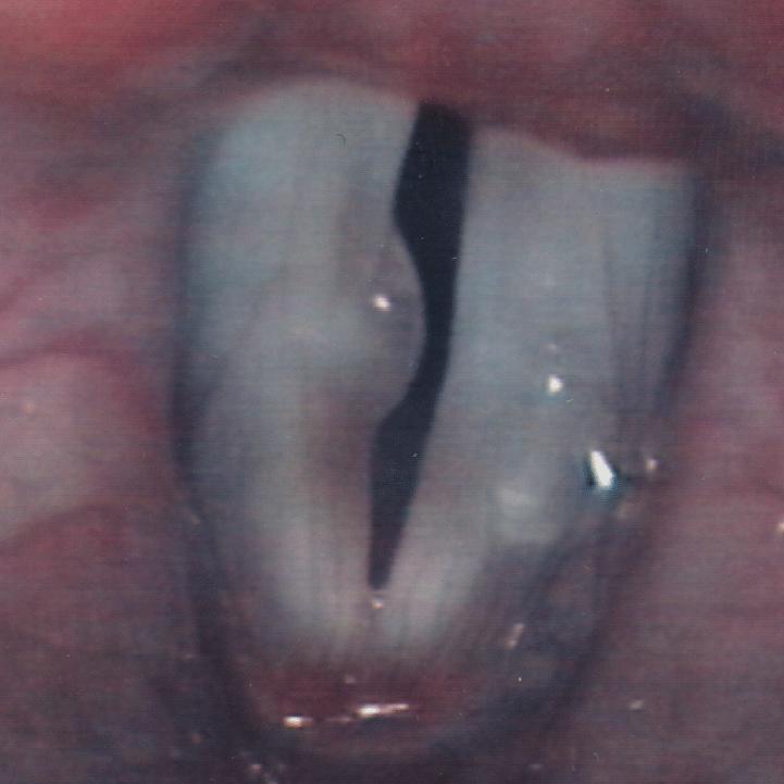
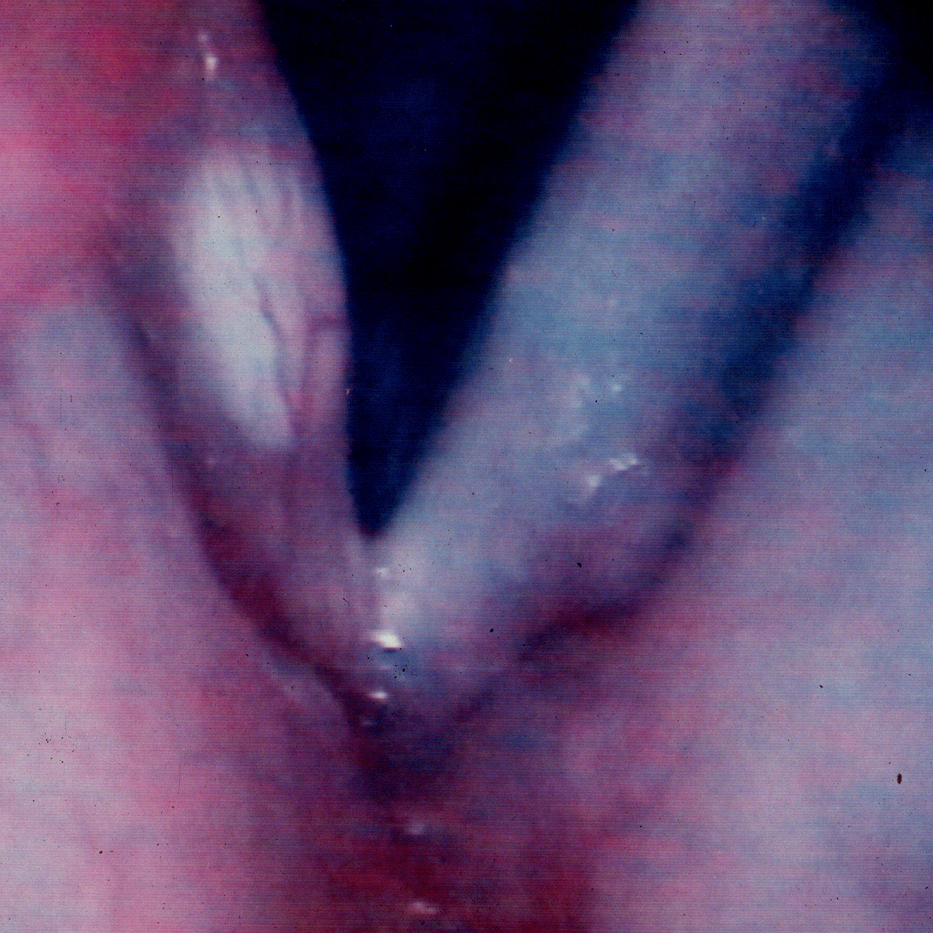
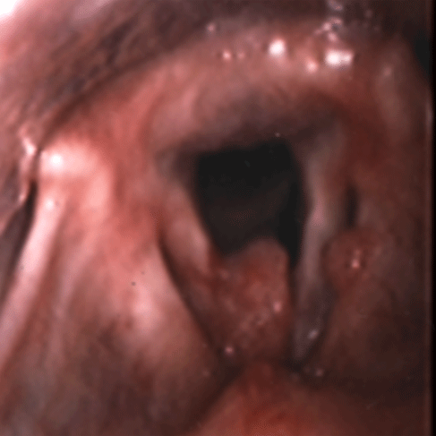
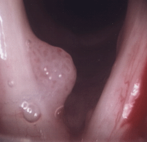
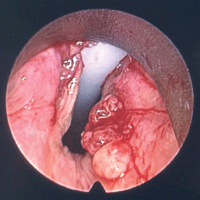
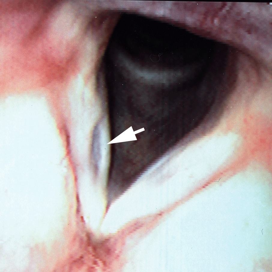
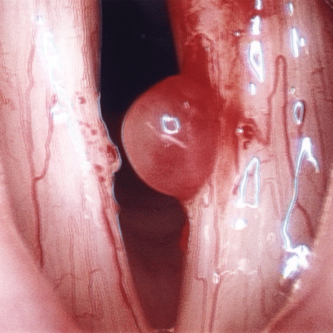
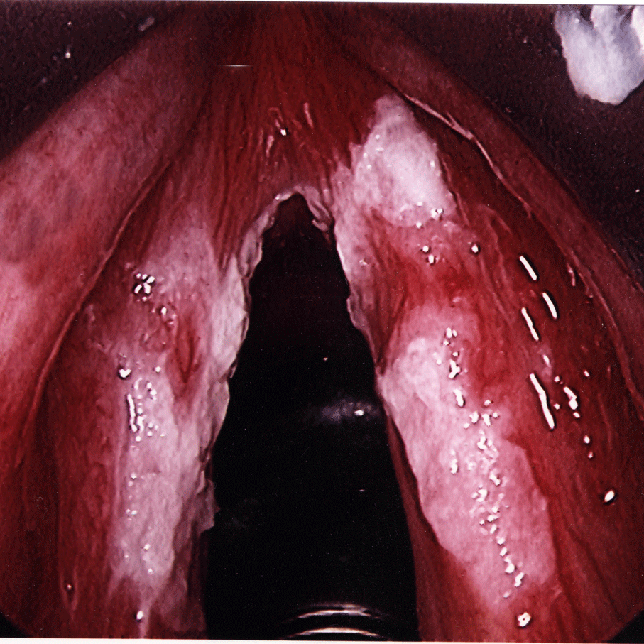
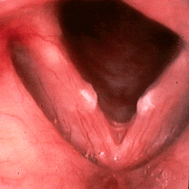
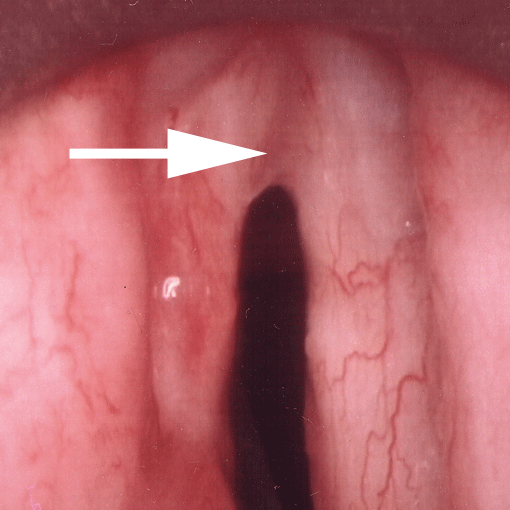
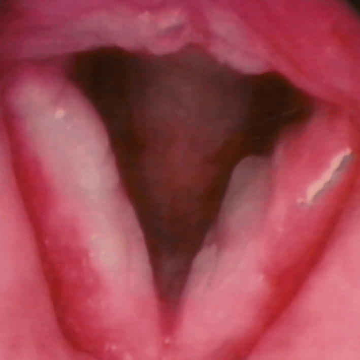
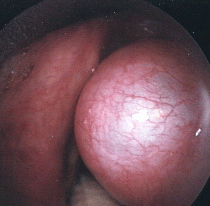
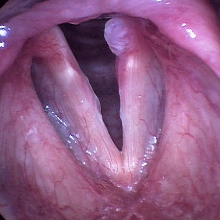
Benign Laryngeal Pathology
Professional Singers
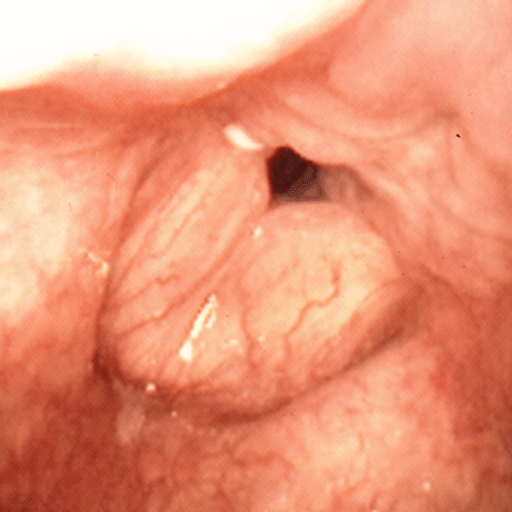
Vocal Fold Paralysis and Paresis
Treatment
Presbylarynx
Spasmodic Dysphonia
Airway Disorders
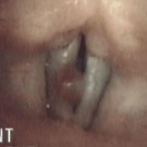
Airway Stenosis
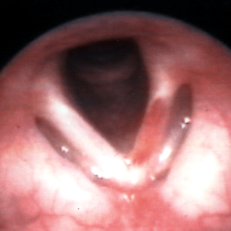
Bilateral Vocal Fold Immobility
Tracheotomy Tube Dependence
Other
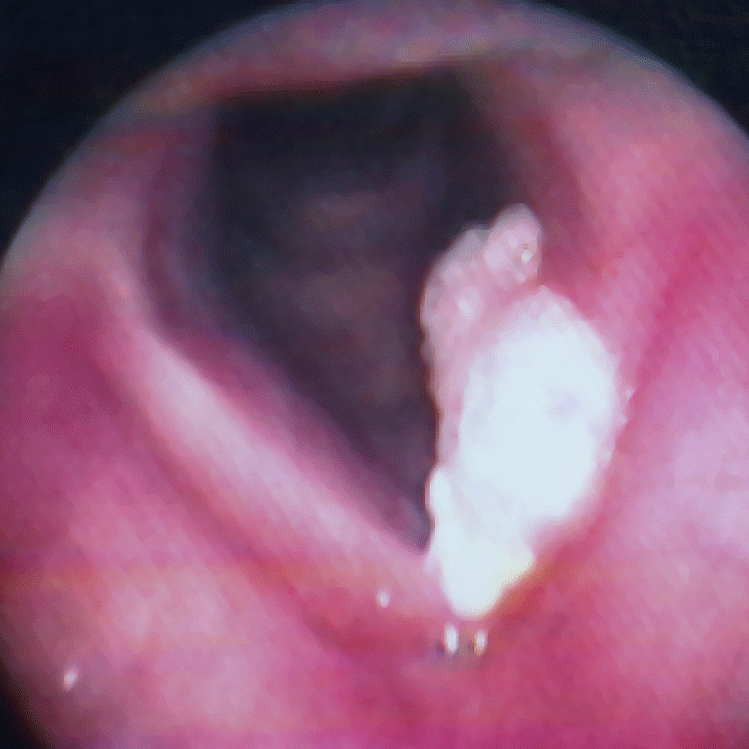
Glottic (vocal cord) Cancer
Extraesophageal Reflux
Reflux Therapy
Chronic Cough
Throat Pain
Services We Offer
Office-Based Procedures
Laser Therapy
Vocal Fold Augmentation
Esophagoscopy
pH Testing
Tracheoesophageal Puncture (TEP)
Bronchoscopy
Comprehensive Swallow Evaluation
Dysphagia Therapy
Comprehensive Voice Evaluation
Voice Therapy
Laryngeal Botulinum Toxin
Voice Restoration after Laryngectomy
About Us
Our Research
Publications
For Our Patients
About Your Procedure
UC Davis MyChart
Resources
Anatomy & Physiology of Swallowing
Voice Anatomy and Physiology
From The Literature
Useful Links
American Speech-Language-Hearing Association
Dysphagia Research Society
NFOSD
NeckBreathers
Spasmodic Dysphonia Association
swallowingdisorders.org
Tracheotomy Info
Oley Foundation
Latest News
Blog
Newsletter Archive
Contact Us
Benign Laryngeal Pathology